Why Do a Urinalysis?

DOWNLOAD this article
Urinalysis is one component of the minimum patient database, used as a screening tool for both healthy and sick pets. In addition to the urinalysis, the minimum database also includes a complete blood count (CBC), serum biochemistry profile, heartworm testing, retroviral testing (for felines), and fecal floatation with centrifugation. Other tests, such as thyroid screening, radiographs, and blood pressure testing may be considered part of the minimum database, depending on the age and health status of the patient. Urine is produced by the kidneys. The functional unit of the kidney is the nephron, which consists of the glomerulus, the filtration unit, and the tubule, which modifies the filtrate. The end product is urine.A urinalysis involves checking the appearance, concentration, and content of urine. It is used to assess the overall health of the kidney and urinary system as well as detect and manage a wide range of disorders, such as urinary tract infections, diabetes, liver and kidney disease. A large amount of information can be obtained from this simple test. Urine can be collected from the patient by cystocentesis, catheterization, or free catch (voided) methods. For routine wellness screening, midstream (voided) collections are adequate if results are negative. Abnormal findings should be confirmed by checking a sterile urine sample obtained by cystocentesis. An early morning sample is recommended. It is also important that the urine sample is fresh. The time a urine sample sits between when it is obtained and when it is analyzed can drastically change the accuracy of the results. This is especially true when evaluating for the presence of crystals. If the sample cannot be examined immediately (within 30 min), keeping the sample refrigerated or chilled (36°–45° F) will help slow the deterioration and minimize bacterial growth. Refrigeration beyond 4 hours leads to diminished value of the sample, as cells begin to lyse and bacteria proliferates. Once refrigerated, the sample should be brought back to room temperature before testing.
EVALUATED URINALYSIS PARAMETERS
- Physical Properties
- Appearance: Color and transparency. For example, a urinary tract infection can make urine look cloudy instead of clear.
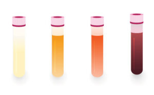
The subjective appearance of the urine sample can provide insights prior to performing objective tests. - Odor: Ammonia, acetone (ketosis) and certain drugs can alter the smell of urine.
- Volume: May indicate polyuria or oliguria.
- Appearance: Color and transparency. For example, a urinary tract infection can make urine look cloudy instead of clear.
- Chemical Examination Chemical testing usually involves inserting a thin strip of plastic known as a dipstick into the urine sample. Chemicals on the stick react with urine and change color to measure the following:
- pH level: The level of acidity can be affected by diet, chemical imbalances, certain metabolic disorders and disease conditions, such as urinary tract infection.
- Nitrites: Can indicate a bacterial infection such as a urinary tract infection.
- Protein:Can indicate primary renal disease, inflammation, and hemorrhage.
- Ketones:Present when the body begins to break down fat as a source of energy. May be a sign of uncontrolled diabetes, diabetic ketoacidosis, or starvation.
- Glucose: Can indicate hyperglycemia, as seen with diabetes mellitus, or stress.
- Bilirubin: A type of waste produced when old red blood cells break down. Bilirubin can indicate liver disease or intravascular hemolysis.
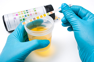
The urine dipstick examination provides an evaluation of the chemical components of the urine sample. - Red and white blood cells: May indicate hemorrhage or infection. A few red and white blood cells can be normal.
- Urine Specific Gravity (USG) Urine Specific Gravity is a measure of renal function and concentrating ability. This value is obtained manually by use of an instrument called a refractometer. Highly concentrated urine can be a sign of dehydration and poorly concentrated urine may be a sign of renal or metabolic disease.
- Microscopic Examination. Traditionally performed by manually examining a drop of urine under the microscope. Prior to analysis, urine must be spun in a centrifuge to concentrate the solid particles and make them easier to examine.
With recent advancements in artificial intelligence (AI) technology, in-house urine sediment analyzers, such as the Heska Element AIM® provide the ability to review a much larger urine sample within minutes. Automation provides a more efficient workflow and eliminates the need for centrifugation.
Microscopic examination on the unstained specimen looks for the following:
- White blood cells: May indicate infection, inflammation, traumatic catheterization or contaminants with free catch samples (urethra and genital system contamination).
- Red blood cells: May indicate renal disease, neoplasia or trauma, possibly associated with cystocentesis or catheterization.
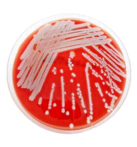
- Bacteria: Can indicate urinary tract infection, or contamination/overgrowth. Rods, cocci, and mixed populations of bacteria are often seen, but the species of bacteria cannot be determined. Culture is required for a definitive classification. Mixed bacteria usually suggests contamination.
- Epithelial (squamous, transitional) cells: May be incidental, but can indicate infection, renal disease, or neoplasia.
- Crystals: May be a sign of kidney stones, metabolic disease or renal toxicity (e.g., ethylene glycol). They may also be artifact or incidental.
- Casts: May be a sign of a tubular damage.
- Other: Fecal parasites, sperm (common in intact males), mold spores, fibers, pollen, starch crystals.
BEYOND THE URINALYSIS
Changes in the urine can be observed before clinical evidence of renal function loss. Based on the urinalysis results, additional testing may be warranted, and early medical intervention can be pursued. The presence of protein in the urine would warrant an evaluation of renal function, including a urine protein to creatinine (UPC) ratio. A urine culture and sensitivity may be pursued if infection is suspected. Stained urine cytology review may be recommended to further classify atypical epithelial cells and to rule out neoplasia. If crystals are present, abdominal imaging may be indicated to rule out urinary stones.
IN SUMMARY
The minimum patient database should be performed as part of any routine wellness examination or when a sick animal is presented. A urinalysis is a quick, safe, and easy screening test that provides invaluable information regarding pet health. Additional tests may be considered depending on urinalysis results and health status of the patient.
REFERENCES
Veterinary Laboratory Medicine Clinical Pathology. Duncan, Prasse and Mahaffey 3rd Ed. pp. 162–174
Veterinary Laboratory Medicine. Meyer, Coles and Rich pp. 71–78
Urinalysis in the dog and cat: A review. Veterinary World. 2020; Vol 13: pp 2133–2141
Learn more about The world’s ONLY fully automated Fecal and Urine Point-of-Care Lab.